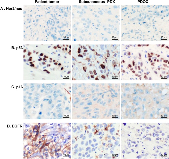
Fig. 2.
Immunohistochemical characterization of the PDOX mouse model of esophageal adenocarcinoma compared to patient tumor. (A) Immunohistochemistry staining for Her2/neu was negative in all tumor cells. (B) p53 demonstrated nuclear staining across all tumor cells. (C) Tumor cells were negative for p16 staining with weak to moderate stromal staining in PDX and PDOX. (D) EGFR membranous staining present in patient tumor cells was lost in PDX and PDOX. Magnification, ×200.

