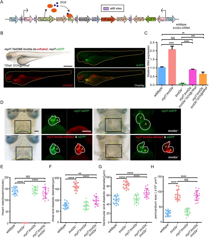
Fig. 2.
Myocardial-specific and DOX-inducible tnnt2a expression fail to rescue heart function in tnnt2a mutant zebrafish. (A) Structure of the tol2-flanked transgenic vector. DOX, doxytetracycline. (B) Lateral views of a 72 hpf transgenic zebrafish after DOX induction at 48 hpf in green and red fluorescence. Scale bar: 300 µm. (C) Real-time RT-PCR of wild-type form of tnnt2a cDNA in wild-type, tnnt2a−/−, Tg(myl7:tetOn; tre:tnnt2a-p2a-mKate2; myl7:eGFP) [Tg(myl7:tnnt2a)] following addition of DOX at 16 hpf, and Tg(myl7:tetOn; tre:tnnt2a-p2A-mKate2; myl7:eGFP; tnnt2a−/−) [Tg(myl7:tnnt2a); tnnt2a−/−] following addition of DOX at 16 or 48 hpf. **P<0.01, ***P<0.001, ****P<0.0001, NS, not significant; n=30 per group. Statistical differences were evaluated using two-way ANOVA. (D) Representative images of hearts of four groups from ventral view. Yellow dotted line, pericardium; white line, heart outline; black box, area of the fluorescent images; V, ventricle; A, atrium. Scale bar: (bright field) 50 µm, (fluorescent) 100 µm. DOX was added at 16 hpf. (E–H) Statistics for heart rate, atrial and ventricular end-diastolic diameter, and pericardium size in four groups; **P<0.01, ****P<0.0001, NS, not significant; n=15 per group. Statistical differences were evaluated using Kruskal–Wallis test (E) or one-way ANOVA (F–H).

