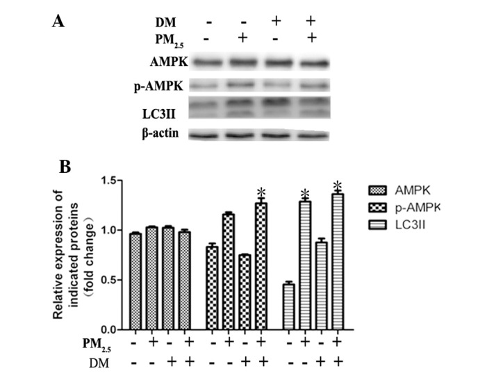
Figure 2.

The AMPK signaling pathway positively regulates PM2.5-mediated autophagy in Beas-2B cells. (A) Western blot analysis of AMPK, p-AMPK and LC3II expression. (B) Quantification relative to β-actin expression. Values are expressed as the mean ± standard deviation of ≥3 separate experiments. Cells were treated with 100 µg/ml PM2.5 for 24 h, following 2 h pre-treatment with the AMPK inhibitor DM (40 µm/l). Dimethyl sulfoxide was tested as a control. PM2.5 enhanced AMPK phosphorylation in Beas-2B cells; however, blocking AMPK activation did not significantly influence PM2.5-mediated autophagy. *P<0.05, vs. control. DM, dorsomorphin; PM2.5, particle matter 2.5; AMPK, adenosine monophosphate-activated protein kinase; p-, phosphorylated; LC3II, microtubule-associated protein 1 light chain 3 II.
