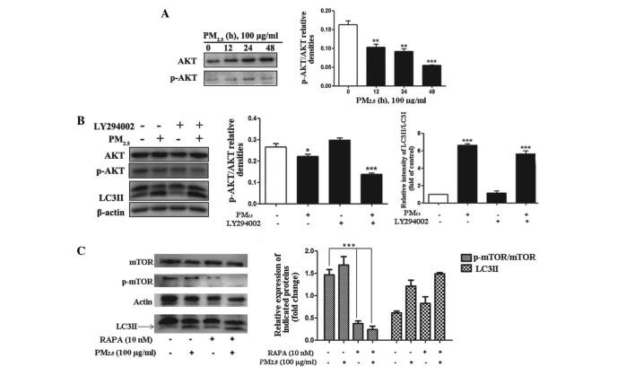
Figure 3.
Role of the PI3K/AKT/mTOR signaling pathways in PM2.5-induced autophagy. Cells were treated with PM2.5 (0, 12, 24 or 48 h) and the expression levels of the indicated proteins were analyzed by immunoblotting. (A) Phosphorylation status of AKT and protein expression levels of AKT were examined by western blot assay in PM2.5-treated Beas-2B cells. (B) Cells were treated with 100 µg/ml PM2.5 for 24 h following 2 h pretreatment with the inhibitor, LY294002. The phosphorylation status of AKT and protein expression level of AKT were analyzed by western blotting in PM2.5-treated Beas-2B cells. (C) Cells were treated with 100 µg/ml PM2.5 for 24 h, following 2 h pre-treatment with rapamycin. The phosphorylation status of mTOR and protein expression levels of mTOR were analyzed by western blotting in PM2.5-treated Beas-2B cells. Values are expressed as the mean ± standard deviation of three independent experiments. *P<0.05, **P<0.01 and ***P<0.001 vs. control. PI3K, phosphatidylinositol 3-kinase; mTOR, mammalian target of rapamycin; PM2.5, particle matter 2.5; p-, phosphorylated; RAPA, rapamycin; LC3II, microtubule-associated protein 1 light chain 3 II.

