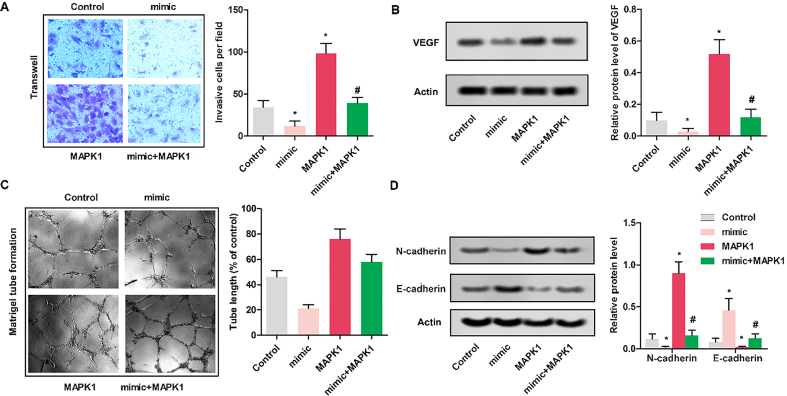
Figure 4.
Up-regulated miR-511 inhibited the invasion of MG63 cells. MG63 cells were randomly divided into 4 groups. Control group: normal MG63 cells; mimic: MG63 cells were transfected with miR-511 mimic; MAPK1 group: MG63 cells were transfected with pcDNA-MAPK1; MAPK1 mimic group: MG63 cells were transfected with pcDNA-MAPK1 and miR-511 mimic. (A) Cell invasion of MG63 was assayed by Transwell assay (magnification, 400). (B&D) The relative protein expressions were detected by western blot in MG63 cells. GAPDH was used as internal control ( 0.05, versus control group; # 0.05, compared with MAPK1 group). (C) Tube formation assay. Photomicrographs were acquired with an inverted microscope (OLYMPUS).

