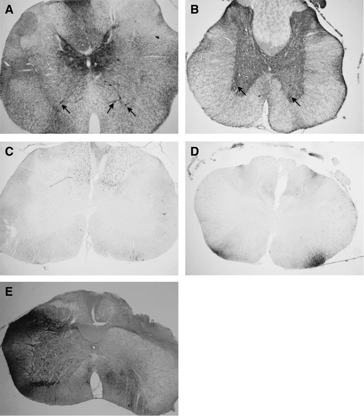
Figure 1.
Gray matter transduction following intrathecal scAAV9 delivery. Seventy-day-old Sprague-Dawley rats received a 30 μL intrathecal injection of scAAV9-CB-GFP via an intrathecal catheter, threaded from the cisterna magna down to lumbar level of the spinal cord. Three weeks later, the spinal cords were harvested, sectioned, and stained for GFP (black stain). (A) Cervical and (B) lumbar spinal cord sections from animals that received the pilot lot from Core A are shown. At all levels of the spinal cord, robust transduction was observed in the gray matter, including cells morphologically similar to motor neurons (arrows). (C) Cervical and (D) lumbar spinal cord sections from animals receiving lot 2 from Core A are shown. In contrast to the animals in (A, B), no gray matter transduction was observed. White matter tracts did stain positively for GFP, possibly due to DRG transduction. (E) When directly injected unilaterally into the spinal cord, a nonpenetrating vector yielded robust transduction of the gray matter on the treated side (left). DRG, dorsal root ganglia; GFP, green fluorescent protein; scAAV9, self-complementary adeno-associated virus serotype 9.

