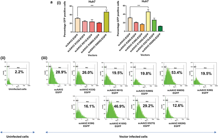
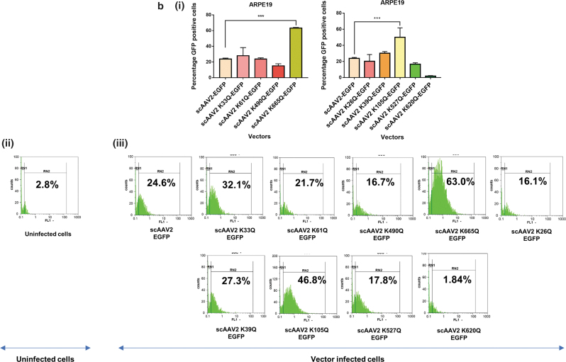
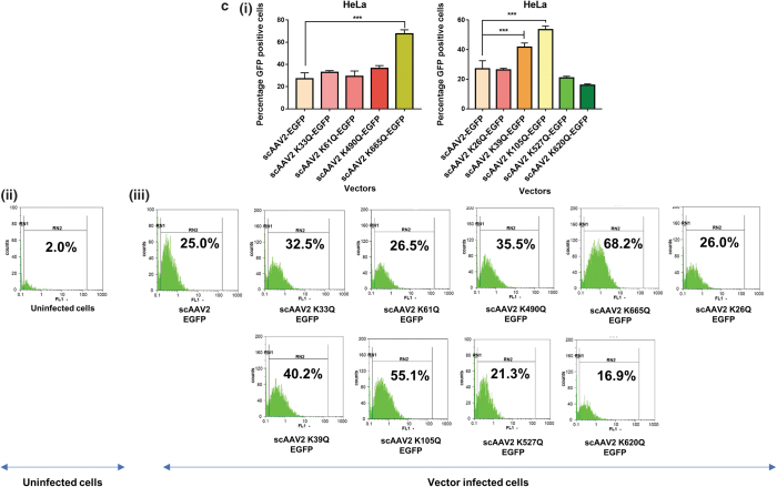
Figure 1.
Transduction efficiency of AAV2 mutant vectors in vitro. About 3 × 10⁴ Huh7, ARPE19, or HeLa cells were mock-infected or infected with scAAV2-EGFP and scAAV2-EGFP mutant vectors for 3 h. Forty-eight hours later, the transgene (GFP) expression was measured by flow cytometry (CyFlow, Sysmex-Partec, Kobe, HP, Japan). Quantitative data for Neddylation or SUMOylation target-site mutants (i) and representative histograms (ii, iii) in Huh7 cells (a), ARPE19 cells (b), or HeLa cells (c) are shown. scAAV2-EGFP control was shared between Neddylation and SUMOylation mutants. Dunnett's multiple comparisons test was used to determine the statistical significance. Data are expressed as mean ± SD, n = 4, ***p ≤ 0.001. Data presented are a representative set from two independent biological replicate analysis. Values on histograms (ii, iii) have been manually shown for one replicate sample from entire analysis. The MFI (mean fluorescence intensity) for the same samples are shown in Supplementary Fig. S3. AAV, adeno-associated virus; ARPE, adult retinal pigmental epithelium; EGFP, enhanced green fluorescent protein; sc, self-complementary. Color images are available online.


