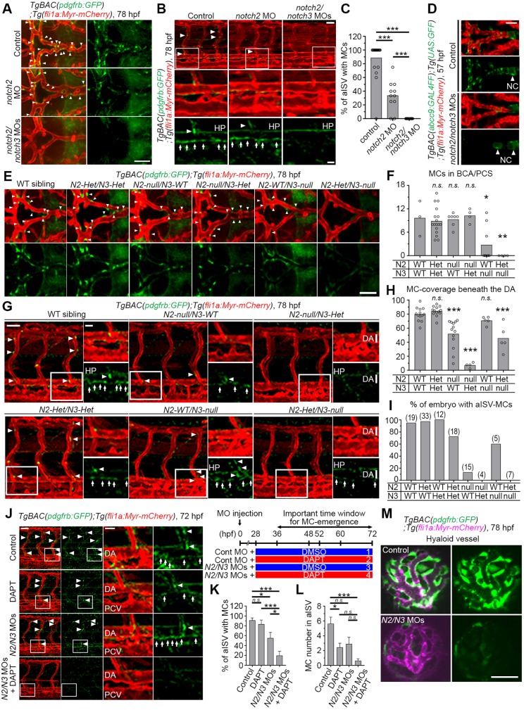
Fig. 3.
Both Notch2 and Notch3 are essential for pdgfrbhigh MC emergence. (A,B) Confocal stack images of the brain (A) or trunk vessels (B) of 78 hpf TgBAC(pdgfrb:GFP);Tg(fli1a:Myr-mCherry) larvae injected with 10 ng control MO, 10 ng notch2 MO or 5 ng each of notch2 and notch3 MOs. (C) The percentage of aISV with pdgfrbhigh MCs, as observed in B (n≥11). (D) Confocal stack images of the brain vessels of 57 hpf TgBAC(abcc9:GAL4FF);Tg(UAS:GFP);Tg(kdrl:DsRed2) embryos injected with 10 ng control MO or 5 ng each of notch2 and notch3 MOs. (E,G) Confocal images of brain (E) and trunk (G) vasculature of the wild-type (WT) siblings or mutants carrying both heterozygous notch2 and notch3 mutations (N2-Het/N3-Het), homozygous notch2 mutation (N2-null/N3-WT), both homozygous notch2 and heterozygous notch3 mutations (N2-null/N3-Het), homozygous notch3 mutation (N2-WT/N3-null) or both heterozygous notch2 and homozygous notch3 mutations (N2-Het/N3-null) with the TgBAC(pdgfrb:GFP);Tg(fli1a:Myr-mCherry) background at 78 hpf. (F,H,I) The number of pdgfrbhigh MCs in BCA and PCS in notch2 and notch3 mutants, as shown in panel E (F), the percentage of the DA with MC coverage (MC-covered/total DA length in observed areas were measured using Imaris software) (H) or the percentage of embryos with pdgfrbhigh MCs in aISV (I) in notch2 and notch3 mutants, as shown in G. The total number of observed larvae are shown on the top of each bar in I. (J) Confocal stack image of the trunk vessels of 72 hpf TgBAC(pdgfrb:GFP);Tg(fli1a:Myr-mCherry) larvae injected with control MO or low dose (0.75 ng each) of notch2 and notch3 MO combined with subsequent treatment with DMSO or low dose (5 µM) of DAPT from 36 to 72 hpf as shown in the scheme to the top right of J. (K,L) The percentage of aISV with pdgfrbhigh MCs (K) and pdgfrbhigh MCs number in aISV on one side of the observed trunk (L), as observed in J. Data are mean±s.e.m. (n≥4). (M) Confocal stack image of the hyaloid vessels of 78 hpf TgBAC(pdgfrb:GFP);Tg(fli1a:Myr-mCherry) larvae injected with 10 ng control MO (top) or 5 ng each of notch2 and notch3 MO (bottom). Boxed regions are enlarged to the bottom or right. Arrows and arrowheads indicate pdgfrbhigh MCs beneath the DA and in ISV/BCA/PCS, respectively. *P<0.05, **P<0.01, ***P<0.001, significant difference between two groups. n.s., not significant. In F and H, significant difference between WT and indicated mutant group. HP, hypochord; NC, notochord; PCV, posterior cardinal vein. Scale bars: 50 µm; 20 µm in enlarged images.

