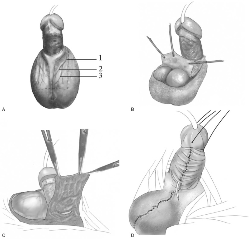
Figure 3.

Schematic illustration of the procedure. (A) The flap, pedicled on the bilateral anterior scrotal artery, was taken from the anterior side of the scrotum. The dotted lines represent the skin incision. The flap was supplied by (1) the anterior scrotal artery, (2) the internal branch of the anterior scrotal artery, and (3) the external branch of the anterior scrotal artery. (B) The flap was partially excised and pulled towards the penis. (C) The flap was reversed and wrapped around the penis from the dorsal side to the ventral side. (D) Primary closure of the donor site.
