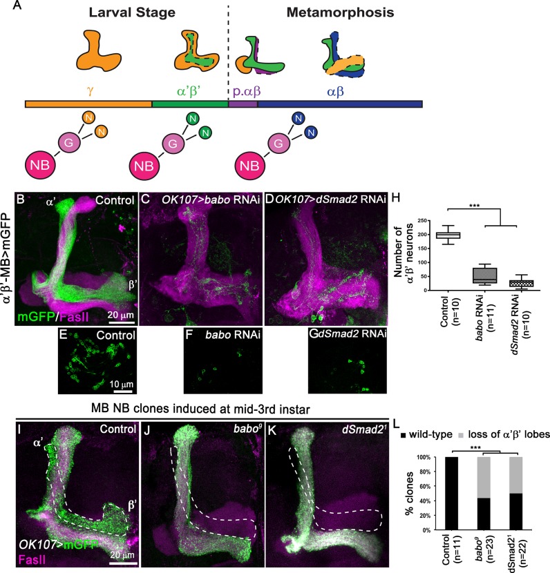
Fig 1. TGF-β signalling controls α’β’ MB neuron fate specification.
(A) Schematic drawings representing the sequential generation of the four distinct subtypes of MB neurons, which can be classified based on their axonal projection patterns (orange, γ neurons; green, α’β’ neurons; purple, pioneer αβ neurons; blue, αβ neurons). MB neuroblasts (NBs) undergo multiple rounds of asymmetric divisions to generate ganglion mother cells (G) that in turn divide to produce two postmitotic neurons (N). (B-D) Adult MB lobes from control (B), OK107>babo RNAi (C) and OK107>dSmad2 RNAi (D) brains stained with anti-FasII antibody (magenta). (E-G) Single confocal section thought the cell body cluster of adult MB neurons from control (E), OK107>babo RNAi (F) and OK107>dSmad2 (G) brains. Green: GMR26E01-LexA-α’β’-MB-driven mGFP (mCD8::GFP) in B-G. (H) Quantification of the number of adult α’β’ MB neuron cell bodies from control, OK107>babo RNAi and OK107>dSmad2 RNAi brains. Statistical comparison to the control: ***, p<0.001 (two tailed t test). (I-K) Adult MB lobes from control (I), babo9 (J) and dSmad21 (K) neuroblast MARCM clones generated at mid-third instar, labelled with mGFP (green) using the GAL4-OK107 driver and stained with anti-FasII antibody (magenta). (L) Quantification of α’β’ MB fate defects in control, babo9 and dSmad21 neuroblast clones. Statistical comparison to the control: ***, p<0.001; (Fisher’s exact test).

