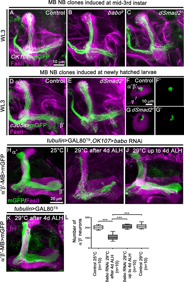
Fig 2. TGF-β signalling is dispensable for the initial α’β’ MB neuron production.
(A-C) WL3 MB lobes from control (A) (n = 8), babo9 (B) (n- = 7) and dSmad21 (C) (n = 12) MARCM NB clones induced at mid-third instar stage, labelled with mGFP (green) using the GAL4-OK107 driver and stained with anti-FasII antibody (magenta). (D, E) WL3 MB lobes from control (D) (n = 5) and dSmad21 (E) (n = 9) MARCM NB clones generated in newly hatched larvae, labelled with mGFP (green) using the α’β’-specific GAL4-c305a driver and stained with anti-FasII antibody (magenta). (F-G’) Cross sections of control (F,F’) and dSmad21 (G,G’) WL3 MB peduncles from neuroblast MARCM clones generated in newly hatched larvae labelled with mGFP (green) using the GAL4-c305a driver and stained anti-FasII antibody (magenta). The arrows indicate the γ axons (FasII-positive area) in the outer layer and the α’β’ axons in the core layer (FasII-negative area). (H, I) Adult MB lobes from tubulin>GAL80TS,OK107>babo RNAi animals raised at 25°C (H) or at 29°C starting from 4d ALH (I). (J) Adult MB lobes from tubulin>GAL80TS,OK107>babo RNAi animals raised at 29°C and switched to 25°C starting from 4d ALH. (K) Adult MB lobes from tubulin>GAL80TS RNAi animals raised at 29°C starting from 4d ALH. The temperature shift is at 4d after larval hatching (ALH) corresponding to the late larval stage. Magenta: anti-FasII staining. Green: GMR26E01-LexA-α’β’-MB-driven mGFP in F,I. (L) Quantification of number of adult α’β’ MB neuron cell bodies from the temperature-dependent babo RNAi experiments in H-K. Statistical comparison to the control: ***, p<0.001 (two tailed t test).

