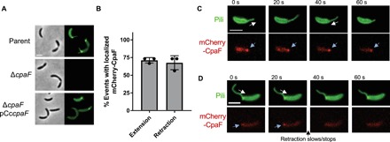
Fig. 2. CpaF is required for tad pilus synthesis and is localized during both pilus extension and retraction.

(A) Representative images of hyperpiliated CB13 pil-cys strains labeled with Alexa Fluor 488 maleimide (AF488-mal). (B) Quantification of pilus extension and retraction events with localized mCherry-CpaF. Data are from 15 extension, and retraction events are from three independent, biological replicates (n = 45 total extension and retraction events). Error bars show means + SD. (C) Representative time-lapse images of mCherry-CpaF localization during both pilus extension and retraction. (D) Representative time-lapse images of mCherry-CpaF delocalization during pilus retraction that correlates with halted retraction. Scale bars, 2 μm. White arrows indicate the direction of pilus movement (away from the cell body is extension, and toward the cell body is retraction), and blue arrows indicate mCherry-CpaF foci.
