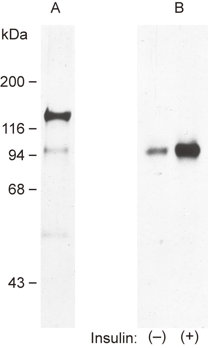
Figure 4.
Phosphorylation of the β subunit of a highly purified insulin receptor preparation. Lane A shows silver staining of the highly purified human receptor after SDS-PAGE under reducing conditions. Lanes B show the incorporation of 32P from [γ-32P]ATP into the highly purified receptor after incubation with or without 0.1 µM insulin for 30 min. The incorporation was detected by SDS-PAGE under reducing conditions followed by autoradiography. Modified with permission from ref. 25.

