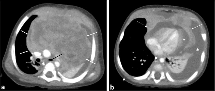
Fig. 10.
An 8-month-old boy with T-cell lymphoblastic lymphoma. a Contrast-enhanced axial CT image of the superior mediastinum and (b) at the level of the cardiac ventricles demonstrate a large heterogeneously enhancing anterior mediastinal mass (white arrows) with pericardial invasion (dashed arrows). There is narrowing of the left main bronchus (black arrow), left lung collapse and a large pericardial and left pleural effusion (asterisks)

