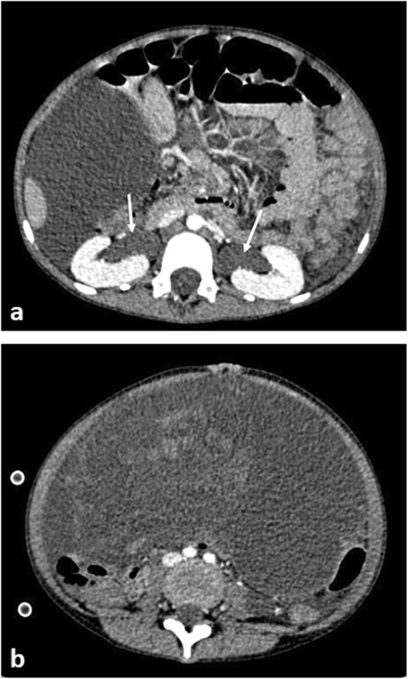
Fig. 17.

An 8-year-old girl with large Sertoli Leydig cell ovarian tumour arising from the right ovary. a Axial contrast-enhanced CT of the upper abdomen demonstrates bilateral hydronephrosis (white arrows) secondary to (b) the large intra-abdominal mass, which was compressing the distal ureters (not visible in image)
