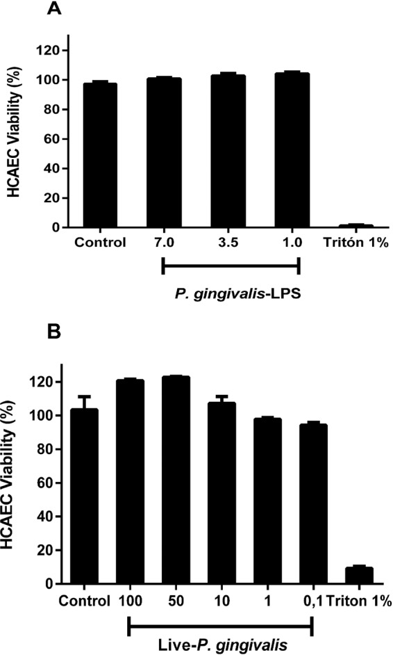
Figure 1.

Viability of HCAECs after repeated treatments with live-P. gingivalis (A) and P. gingivalis-LPS (B). The HCAECs were stimulated to repeated live-P. gingivalis (MOI 1:100 - 1:0,1) and P. gingivalis-LPS (1.0, 3.5 and 7.0 µg/mL) exposures, during 24 h. Cell viability was determined according to the fluorometric detection after reduction of resazurin in the resorufin product using AlamarBlue. 1% was considered our positive control of cell death. Percentage of cell viability with respect to the control. *Represents the statistical difference with respect to the control or without stimulus. (p < 0.05). Three independent experiments were performed; the results are presented as the means ± SEM (n = 3).
