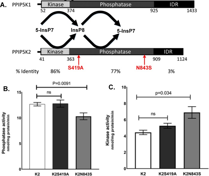
Figure 2.
Schematic diagram of PPIP5K1 and PPIP5K2 domains and the in vitro biochemical assays for the phosphate and the kinase activities carrying the identified pathogenic variants. (A) Diagram of PPIP5K1 and PPIP5K2 functional domains with the identified pathogenic variants. The text with arrows shows our identified pathogenic variants located in the phosphatase domain of PPIP5K2. (B) PPIP5K2-phosphatase activity level with the two identified variants. (C) PPIP5K2-kinase activity level with the two identified variants. K2 represents hsa-PPIP5K2, K2S419A represents hsa-PPIP5K2 with Serine 419 to Alanine change, and K2N843S represents hsa-PPIP5K2 with Asparagine 843 to Serine change. Each bar represents four separate independent enzymatic assays.

