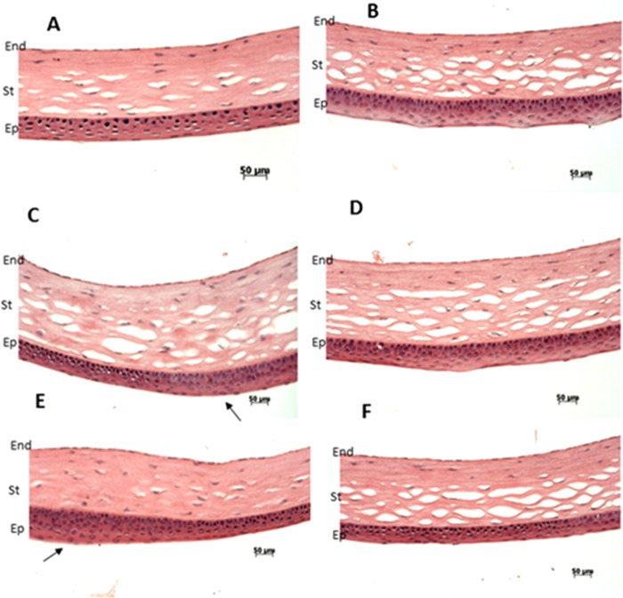
Figure 6.
H&E staining of the mouse corneas. Normal epithelium was observed in wild-type Ppip5k2+/+ littermate controls (A,B). Thickened corneal epithelium layer was observed in some Ppip5k2+/K^ heterozygous (C) and Ppip5k2K^/K^ homozygous (E) mouse corneas. These corneas (C,E) were found to have irregular anterior corneal surfaces using SD-OCT scanning. Normal corneal epithelium thickness was noticed in other Ppip5k2+/K^ heterozygous (D) and Ppip5k2K^/K^ homozygous (F) mouse corneas. These corneas (D,F) were found to have no irregular anterior corneal surfaces using OCT scanning at 3 months of age. The black arrows showed the thickened epithelium in panels C and E. Abbreviations: Ep for epithelium, St for stroma, and End for endothelium of the mouse cornea.

