Figure 1.

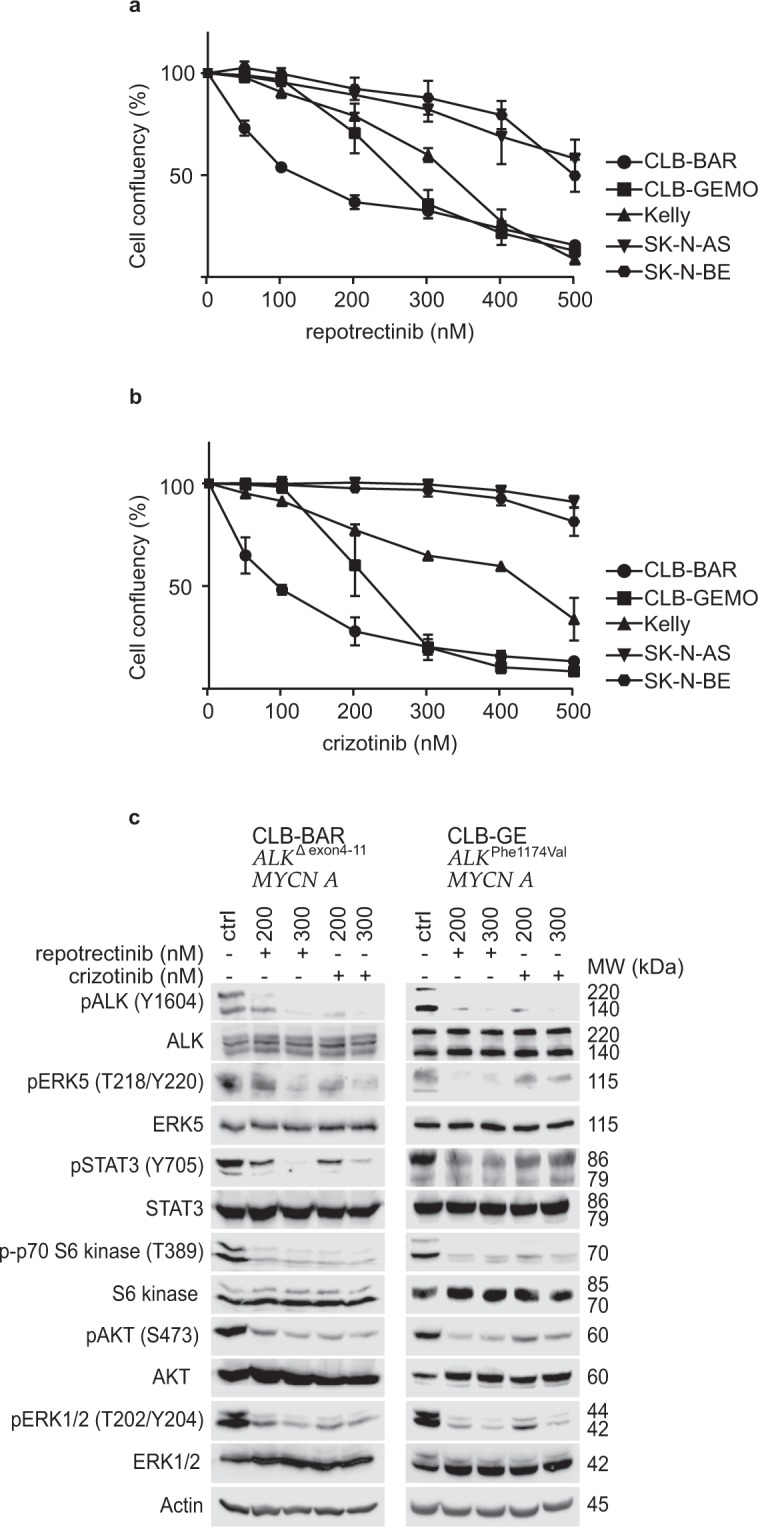
Repotrectinib inhibits proliferation of neuroblastoma cells. Neuroblastoma cells were treated with either repotrectinib or crizotinib for five days, and cell confluency monitored by live cell imaging. (a) Repotrectinib response curve and (b) crizotinib response curve. The plotted values are means ± S.D. from growth curves from three independent experiments performed in triplicate. (c) CLB-BAR and CLB-GE, both ALK-addicted cell lines, were treated with either 200 or 300 nM reptorectinib. Crizoitinib (200 nM and 300 nM) was employed as a positive control. Cell lysates were resolved on SDS/PAGE followed by immunoblotting for pALK (Y1604), pAKT (S473), pERK 1/2(T202/Y204), pERK5 (T218/Y220), p70 S6 kinase (T389) and pSTAT3 (Y705). Total protein for ALK, AKT, ERK 1/2, ERK5, S6 kinase and STAT3 were used as protein loading controls. Phosphorylated proteins were visualized prior to stripping and immunoblotting for total protein. CLB-BAR cells have a genomic deletion in ALK between exon 4–11, resulting in an ALK band of approximately 170 kDa54. The CLB-GE cell line expresses a mutant full-length version of ALK-F1174V, which is cleaved resulting in the detection of two bands with the antibody employed here. Blots are representative of three independent experiments.
