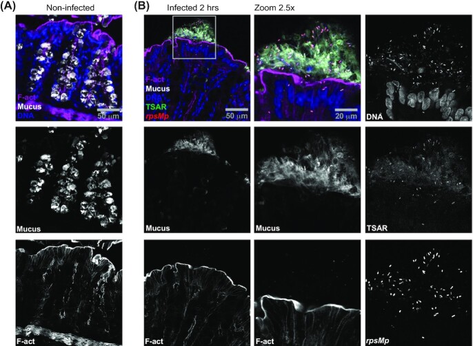
Figure 1.
Massive mucus release of the colonic mucosa is induced by luminal S. flexneri displaying low T3SA activity. Colonic tissue sections infected or not with WT Shigella flexneri harboring pTSAR1.1 and labeled with Wheat Germ Agglutinin (WGA), phalloidin and DAPI were imaged using confocal microscopy. A micrograph overlay of a non-infected tissue section (A). A micrograph overlay of an infected tissue section at 2 h post-challenge (left panel). The indicated channels corresponding to the boxed area are magnified by 2.5-fold and represented in the central and right panels (B). Sections are positioned with the lumen on top and are representative of infection foci observed in two animals.

