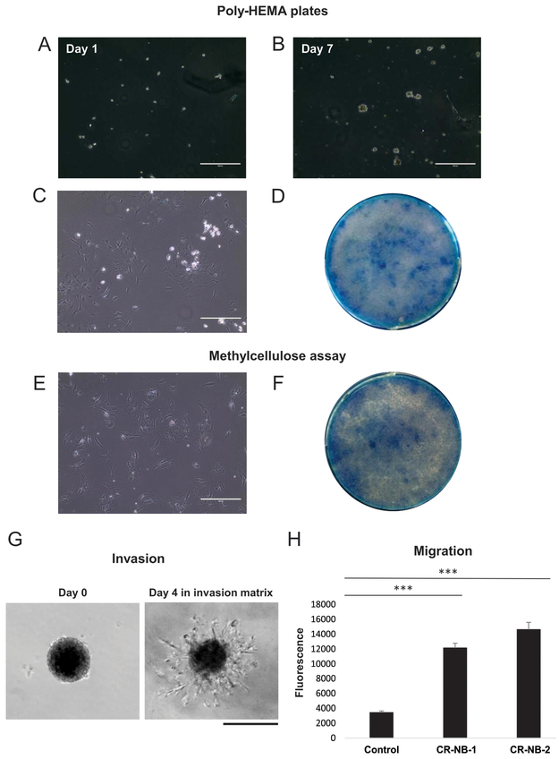
Figure 7. Malignant features of CR-NB cell lines.
A-B. Subsequent stages of the anchorage-independent growth of CR-NB cells on poly-HEMA low attachment plates. C-D. Cells grown in suspension on poly-HEMA plates for 7 days were plated into standard cell culture plates under established optimal culture conditions (C) and upon reaching confluency visualized by methylene blue staining (D). E-F. Cells cultured for 7 days under anchorage-independent conditions in methylcellulose were plated into the standard culture plates and their growth assessed as above. Scale bars represent 400 μm. G. CR-NB cells invade the extracellular matrix 4 days after embedding. Scale bar represents 200 μm H. CR-NB cell lines showed a significant motility in a transwell migration assay. Mean fluorescence intensity ± standard error is shown. *** represent p<0.001 as compared to control (no cells).

