Figure 1.
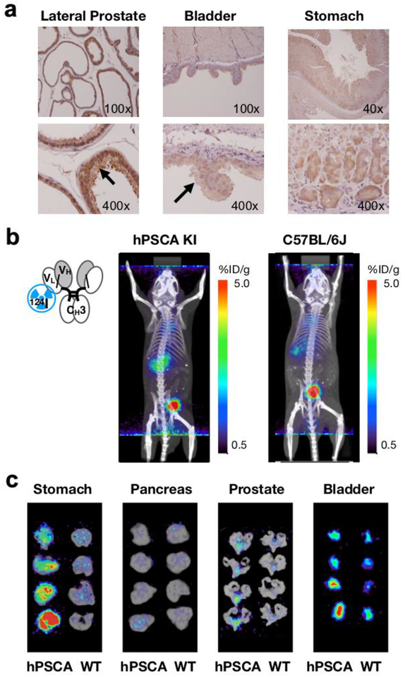
hPSCA-expression in hPSCA KI mice and [124I]A11 Mb immunoPET. a Human PSCA knock-in mice (hPSCA KI × C57BL/6J) show low level expression of PSCA in the normal prostate, bladder and stomach similar to the expression pattern seen in humans. Immunohistochemical staining was performed with anti-hPSCA mouse mAb 1G8 (parental antibody of A11 Mb and A2cDb). b [124I]A11 Mb (4.44 MBq/30 μg) was injected into hPSCA KI and wildtype C57BL/6J mice (n = 4 each) and immunoPET scans were acquired 20 h post injection. c Ex vivo PET/CT scans of normal PSCA-expressing tissues. hPSCA KI mice (left panels) showed slightly higher tracer uptake in the stomach and the bladder compared with C57BL/6J wild type mice (right panels).

