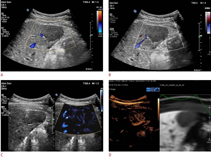
Fig. 2. Representative example of a MicroFlow Imaging examination in a 56-year-old male patient with hypervascular hepatocellular carcinoma.
A 2.8-cm hypoechoic lesion is detected in the right medial section of the liver. A, B. On color Doppler imaging (A) and power Doppler imaging (B), this tumor does not show hypervascularity. C, D. The MicroFlow Imaging examination shows a mixed pattern of vascularity, combining the basket and vessels-in-tumor patterns (C), which is confirmed on a contrast-enhanced ultrasound examination (D).

