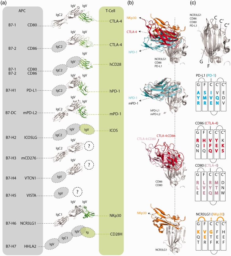
Figure 2.
Structural analysis of B7 and CD28 members. (a) Structures of select B7 (gray) and CD28 (green) members. From top to bottom PDB codes: 1I8L (hCD80:hCTLA4), 1I85 (hCD86:hCTLA4), 1YJD (hCD28), 4ZQK (hPD-1:hPD-L1), 3BP5 (mPD-1:mPD-L2), 4I0K (mCD276 has two Ig domains; hCD276 has four), 4GOS (hVTCN1), 3PV6 (hNCR3LG1:NKp30). (b) Comparing IgV domains interaction angle for 4ZQK (hPD-1:hPD-L1), 3BP5 (mPD-1:mPD-L2), 1I8L (hCD80:hCTLA4), 1I85 (hCD86:hCTLA4), and 3PV6 (hNCR3LG1:NKp30). (c) Beta sheets of aligned B7 members at the counter receptor interface, and specific residues that structurally align in 3D space. Residues and loops that are in contact with the CD28 members are colored and in bold.

