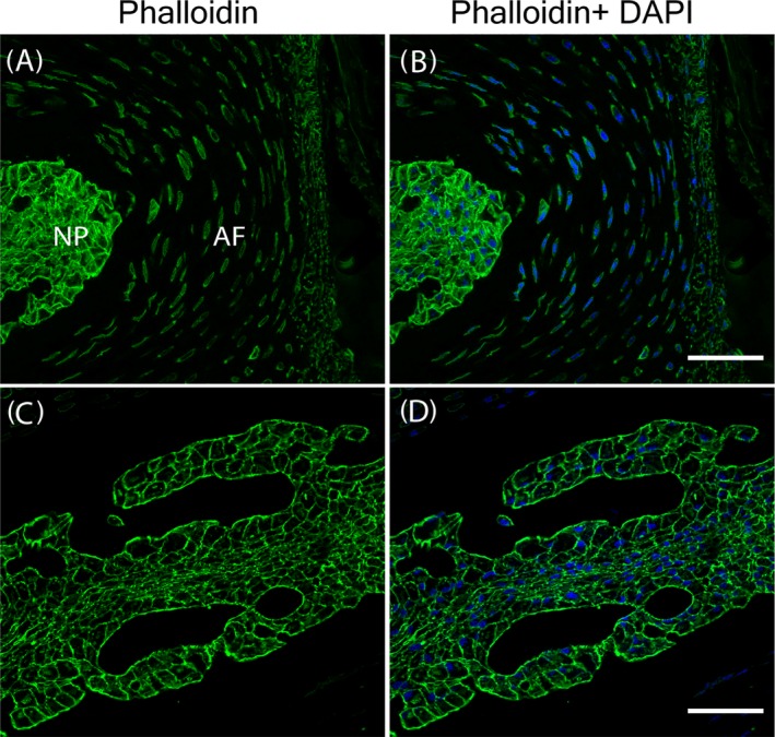
Figure 5.

F‐actin aligns along the cell membrane in the mouse lumbar spine intervertebral disc (IVD). A,B, Low magnification images of lumbar spine IVD. C,D, High magnification images of the nucleus pulposus (NP). Scale bars: 250 μm for the low magnification images and 25 μm for the high magnification images
