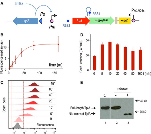
Figure 4. Evaluation of the functionality of the digitalizer module.

- Schematic representation of the relevant features of the specific regulatory device used to evaluate the efficiency of the digitalizer module by means of a reporter msf•GFP gene.
- GFP was used as fluorescence reporter to analyse the OFF and the ON expression profiles of the digitalized version of XylS/Pm system in flow cytometry experiments. The plot represents the median and the SD values of fluorescence from three independent experiments without inducer (t = 0) and upon 3MBz induction along time, at the indicated time points.
- Representative experiment showing the distribution of fluorescence in cell population under non‐inducing conditions (t = 0) and after induction at the indicated time points (t = 5 min to t = 160 min), as indicated. The region considered as negative for the fluorescence signal is marked between red dashed lines, as assessed by control cells carrying a plasmid with a promoterless GFP (grey plot).
- Analysis of the population with respect to cell‐to‐cell heterogeneity by means of the coefficient of variation (percentage CV*100) at the off state (t = 0) and along the induction of the system, as described. Data correspond to the mean and SD values obtained from three separate experiments.
- The NIa protease was used as a sensitive reporter to test both the basal and the induced activity driven by the digitalized XylS/Pm device. The Western blot shows the cleavage of a NIa‐sensitive TpiA target protein under the indicated conditions, which are equivalent as those used for the experiment presented in Fig 2. The control lane #1 was pasted from a different gel to show the location of the intact protein (see a detailed cleavage kinetics in Appendix Fig S6).
