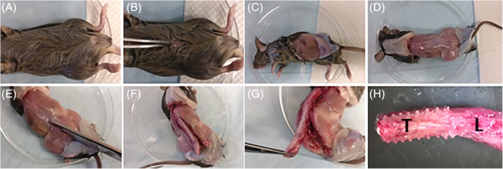
Figure 1.

Spinal column isolation. A, Douse the fur with 70% ethanol to prevent contamination and limit spread of hair. Place the mouse on its back. B, An incision is made at level of the umbilicus on the ventral skin. C, A transverse tear is made either manually or with scissors in both directions from the incision to the dorsal spine. D, The pelt is pulled to the base of the tail and to the base of the neck. The spinous processes should now be visible, allowing visualization of the boundaries of the spine. E, With scissors, begin excising the spine from its lateral attachments of muscle, bone, and fascia in a parallel position to the spine. The process is easiest if beginning at the base of the tail and working in a superior direction, ending distal to the region of interest. F, Once one side has been cut, proceed to the opposite side, cutting in the same areas. G, Once the spine has been freed from its lateral attachments, it is still connected both superiorly and inferiorly to the cervical and sacral spine and anteriorly by viscera. Make a transverse cut below the attachment of the femurs, and, pulling the spine away from the body, begin cutting the anterior attachments. Proceed superiorly until the regions of interest have been detached anteriorly, and make another transverse cut through the superior attachment, freeing the spinal column from the body. H, The excised spine. L, lumbar region; T, thoracic region
