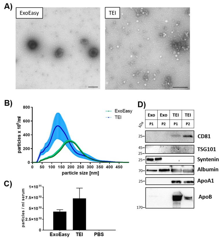
Figure 1.
Characterization of extracellular vesicles. EVs were isolated by either membrane affinity (ExoEasy) or precipitation (TEI) methods. (A) Transmission electron microscopy was utilized to analyze morphology of isolated particles; scale bar = 200 nm. (B) The size profile of particles was assessed by nanoparticle tracking analysis (NTA) (mean and SEM of n = 4 individuals). (C) The total amount of particles from NTA in (B) was calculated. (D) Western blot analysis determined the presence of EV-associated markers and contamination with lipoproteins and soluble proteins. Serum from two healthy individuals (P1, P2) was used for EV isolation. A total of 20 µg total protein was loaded onto the gel.

