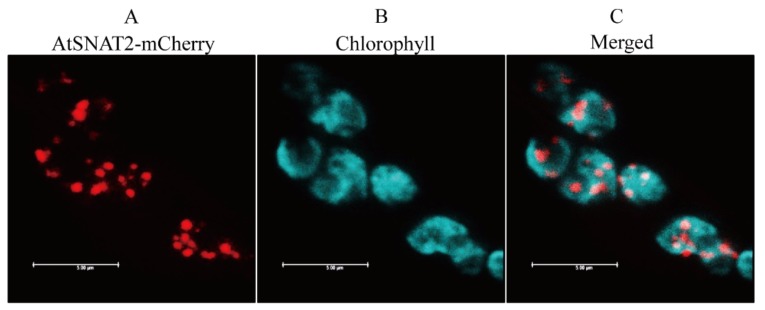
Figure 2.
Subcellular localization of AtSNAT2. (A) Red fluorescence of AtSNAT2-mCherry. (B) Cyan fluorescence of chlorophyll. (C) Merged fluorescent images (A+B). Tobacco leaves were infiltrated with Agrobacterium tumefaciens GV2260 expressing the XVG-inducible AtSNAT2-mCherry fusion protein. Bars: 5 μm.

