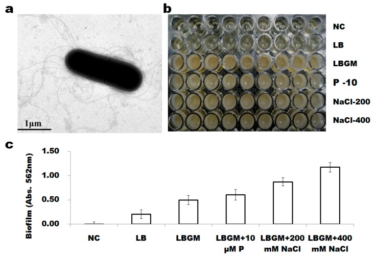
Figure 3.
The morphology of B. altitudinis WR10 and its biofilm formation: (a). transmission electron microscope image of WR10; (b). phenotypes of WR10 biofilm formation in polystyrene microplates under static incubation grown in different media; (c). quantification of biofilms of WR10 by crystal violet staining. Data were mean of three independent experiments. NC, negative control, LB medium without inoculation of bacterium; LB, WR10 grown in Luria-Bertani medium (LB); LBGM, WR10 grown in LB supplemented with 1% of glycerol and 200 μM MnSO4; P–10, WR10 grown in LBGM + 10 μM KH2PO3; NaCl–200, WR10 grown in LBGM + 200 mM NaCl; NaCl–400, WR10 grown in LBGM + 400 mM NaCl.

