Figure 5.
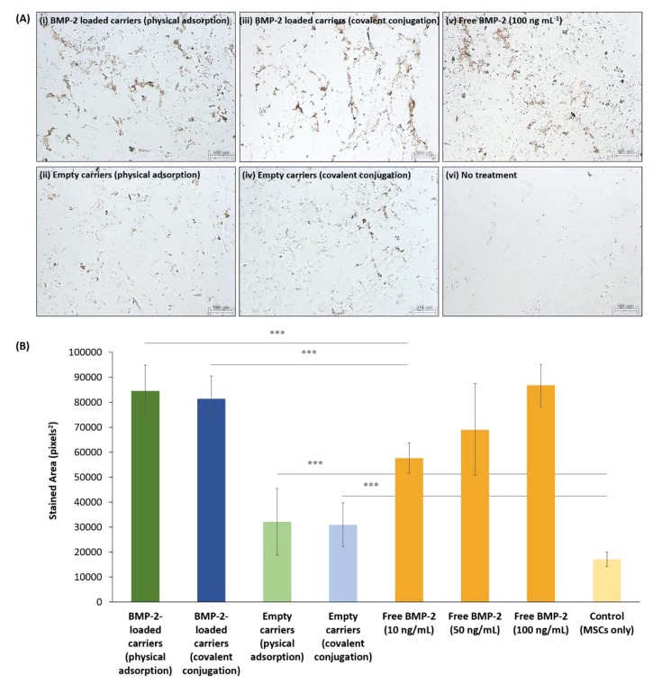
ALP staining of differentiated BM-MSCs after 14 days. (A) Representative light micrographs of cells exposed to (i) BMP-2-loaded PSiO2 carriers via physical adsorption, (ii) Empty PSiO2 carriers, (iii) BMP-2-loaded PSiO2 carriers via covalent conjugation, (iv) Empty chemically-modified PSiO2 carriers, (v) Free BMP-2 solution (100 ng mL−1) and (vi) no treatment (control, cells only). Scale bar = 100 µm. (B) Quantitative analysis of ALP activity, expressed as the average positively stained areas for each condition tested, *** indicates p ≤ 0.005.

