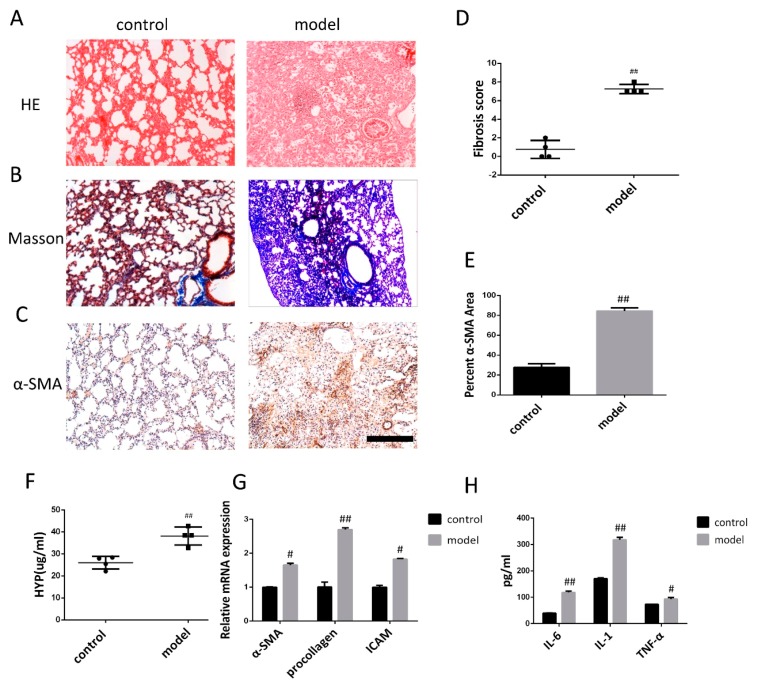
Figure 2.
Pathological changes in lung tissue and the level of inflammatory cytokine in model of mice after bleomycin-induced pulmonary fibrosis formation. (A) The lung section from the mice that have undergone different treatments were stained by hematoxylin–eosin (HE) (n = 4 per group). (B) Lung sections from various treatment groups were subjected to Masson trichrome staining (n = 4 per group). (C) The expression of α-SMA was determined by using immunohistochemical analysis. Scale bar = 200 μm for each picture (original magnification: ×100, n = 4 per group). (D) The lung fibrosis score based on the scoring from Ashcroft score. (E) Quantitative analysis of α-SMA positive area. (F) Hydroxyproline concentration in lung tissue was detected by using alkaline hydrolysis (n = 4 per group). (G) Change of α-SMA, procollagen, and ICAM transcripts were analyzed by qRT-PCR (n = 6 per group). (H) The expression of IL-6, IL-1, and TNF-α was examined by using ELISA in the lung and serum. All data are expressed as the mean ± SD. # p < 0.05, ## p < 0.01 versus control.

