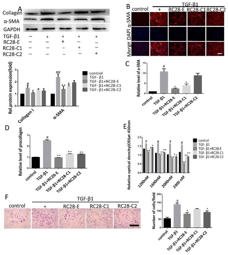
Figure 6.
RC28-E attenuates TGF-β-induced fibroblast activation. (A–D) Human skin fibroblasts line Detroit 551 were treated with RC28-E (2400 nM) or 2400 nM RC28-C1 (VEGF-trap) and RC28-C2 (FGF-trap) in the presence hTGF-β1 (10 ng/mL) for 72 h. (A) The protein expression levels of α-SMA and collagen I were examined by western blot and GAPDH as loading control. Quantitative data were from western blot analysis (n = 3). (B) The expression of α-SMA was detected by immunofluorescence assay. Nuclei were stained with DAPI (blue). Scale bar = 275 μm for each picture (original magnification: ×200). (C,D) Changes of α-SMA and procollagen transcripts were analyzed by qRT-PCR. (E) cells were treated with 1200, 1600, 2000, and 2400 nM RC28-E or 1200, 1600, 2000, and 2400 nM RC28-C1 (VEGF-trap) or 1200, 1600, 2000, and 2400 nM RC28-C2 (FGF-trap), with hTGF-β1 (10 ng/mL) as the control, for 48 h. Relative cell proliferation was determined by using cell counting kit-8. (F) Transwell was used to migration analysis, the migrated cells were stained with crystal violet. Results are shown as mean ± SD. * p < 0.05, ** p < 0.01 versus hTGF-β1 alone. # p < 0.05, ## p < 0.05 versus control (n = 3).

