Figure 3.
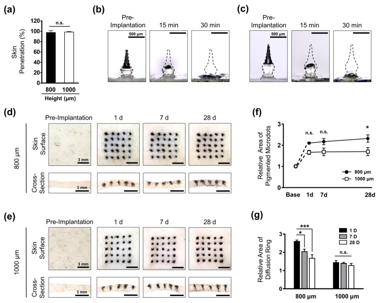
Transcutaneous analysis of micro-pigment-encapsulated biodegradable microneedles (PBMs). (a) Both 800 μm- and 1000 μm-long PBMs showed a high skin penetration success rate. (b) The micro-pigment-encapsulated top layer in both the 800 μm-long PBM and (c) 1000 μm-long PBMs were dissolved at 15 min post-implantation. At 30 min, the unpigmented primary layer was completely dissolved. (d) Implantation of 800 μm-long PBM into the pig cadaver skin resulted in the formation of a large diffusion ring on the skin surface (upper panels). The cross-section of skin confirmed the localization of the micro-pigment up to 28 days (lower panels). (e) The diffusion ring in the 1000 μm-long PBM-implanted skin was barely visible. The dimension of microdots was highly maintained up to 28 days (upper panels). Cross-section images revealed a sharper localization of micro-pigments compared with the 800 μm-long PBM-implanted skin. (f) Comparison of microdots confirmed the well-maintained micro-pigmentation in the 1000 μm-long PBM-implanted skin up to 28 days. (e) The diffusion ring was significantly reduced over time in the 800 μm-long PBM-implanted skin. Data in (a,f,g) are expressed as the mean ± SEM. * p < 0.05, and *** p < 0.001.

