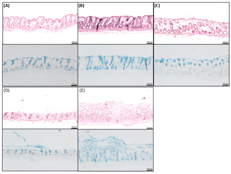
Figure 6.
Representative histology of colonic tissue mucosae after 120 min exposure to sucrose laurate (SL) (1.5 mM, 5 mM, and 10 mM). H & E staining (upper panels, (A–E)) and neutral red and alcian blue staining (lower panels). Bar = 100 μm. (A) KH control; (B) 1.5 mM SL; (C) 5 mM SL; (D) 10 mM SL; (E) 10 mM C10.

