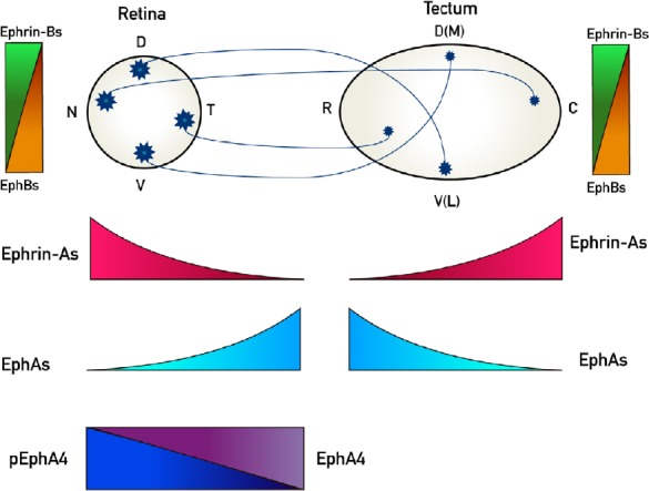
Figure 1.

Representation of retinotectal/collicular projections and the expression patterns of EphAs and ephrin-As along its rostro-caudal axis and the EphBs and ephrin-Bs along its dorso-ventral axis.
Retinal ganglion cells (RGCs) project to contralateral tectum in chicks whereas they project bilaterally to colliculus in mice. Nasal (N) RGCs axons project to caudal (C) tectum/colliculus whereas temporal (T) RGC axons project to rostral (R) tectum/colliculus. Dorsal (D) RGCs axons project to ventral(V)-lateral (L) tectum/colliculus meanwhile ventral (V) RGC axons project to dorsal (D)-medial (M) tectum/colliculus. Ephs and ephrins are expressed in gradients both in the retina and the tectum/colliculus. EphAs (3, 5, 6) (light blue) are expressed in an increasing naso-temporal gradient in the retina, whereas EphA4 presents an even expression along the retina (purple), but it presents a decreasing nasodorsal to temporoventral gradient of phosphorylation (p) (blue). EphAs (3, 6, 7) are expressed in a decreasing rostro-caudal gradient in the tectum/colliculus. Ephrin-As (2, 5, 6) are expressed in a decreasing naso-temporal gradient in the retina and in an increasing rostro-caudal gradient in the tectum/colliculus (red). EphBs are expressed in increasing dorso-ventral gradients both in the retina and the tectum/colliculus (orange) whereas ephrin-Bs are expressed in-decreasing dorso-ventral gradients both in the retina and the tectum/colliculus (green).
