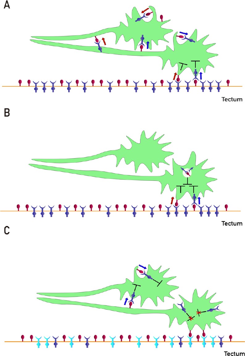
Figure 4.

Different ways of interactions between EphAs and ephrin-As participate in retinotectal/collicular mapping.
(A) Proposed mechanisms of fiber-fiber (FF)- and fiber-target (FT)-chemoaffinity. Segregation of fibers is mediated by FF EphAs (blue)/ephrin-As (red) forward (blue arrow) and reverse (red arrow) signaling besides FT EphAs (blue)/ephrin-As (red) forward (blue arrow) and reverse (red arrow) signaling. Cis forward (blue arrow) and reverse (red arrow) signaling and inhibition (T-shaped symbol) are also shown. The guidance potential is calculated by adding all instantaneous forward and reverse signals impinging on the axons and balancing the sums. The termination zones (TZs) are formed when the guidance potential is zero. (B) Proposed mechanism of co-adaptation. EphAs (blue) and ephrin-As (red), located in membrane lipid microdomains signal in trans forward (blue arrows) and reverse (red arrows) eliciting repulsion. Dispersed, unbound sensors are constitutively endocytosed and might increase cis signaling upon internalization, as anti-parallel orientation is favored in high-curvature membrane vesicles. Enhanced cis signals desensitize growth cones (GCs) to trans signals. Sensitivity returns with the recycling of unbound sensors. (C) Regulation of ephrin-As-dependent EphA4 forward signaling by tectal EphA3 during retinotectal mapping. Ephrin-As (red)-dependent EphA4 (blue) forward signaling (blue arrows) decreases the nasal retinal ganglion cells (RGCs) axon growth before reaching the tectum. Tectal EphA3 (light blue) binds and displaces axonal ephrin-As from axonal EphA4 decreasing its forward signaling and stimulating nasal RGC axon growth toward the caudal tectum and inhibiting TZ formation in the rostral tectum.
