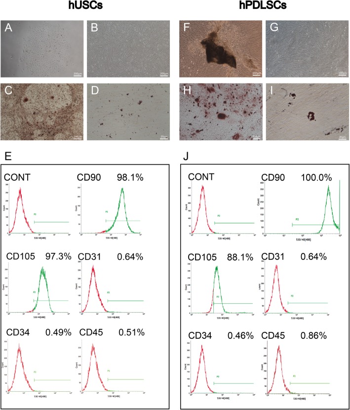
Fig. 1.
Characterization of hUSCs and hPDLSCs. a, f Primary hUSCs and hPDLSCs. b, g hUSCs and hPDLSCs grown in culture medium and exhibiting rice grain-like and long spindle-shaped morphology, respectively. c, h After osteogenic induction for 21 days, mineral deposits formed by hUSCs and hPDLSCs were stained with alizarin red. d, i Cultured in adipogenic medium for 21 days, lipid-laden lobules formed by hUSCs and hPDLSCs were detected with Oil Red O. e, j Cytometric flow analysis indicated that both hUSCs and hPDLSCs expressed the mesenchymal associated markers CD90 and CD105, and were negative for CD31, CD34, and CD45

