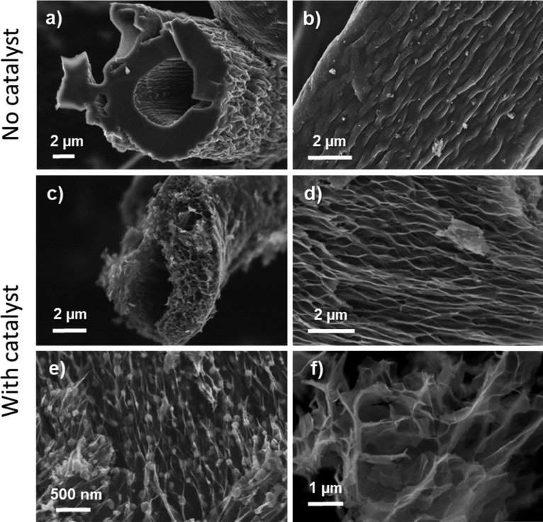
Figure 2.
Representative SEM micrographs: (a, b) Front and side views of a representative fiber from conventional, nontreated MDF carbon carbonized at 300 °C; (c, d) front and side views of a representative fiber of MDF Ni H2O 300 °C sample; (e) MDF Ni H2O 1000 °C sample where globular nickel nanoparticles are appreciable under light contrast before acid washing, and (f) MDF Ni H2O 1000 °C after acid washing with HCl.

