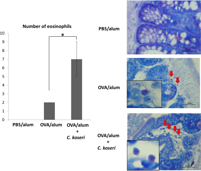
FIG 5.
Numbers of eosinophils that infiltrated the small intestines of ovalbumin (OVA)/alum mice after the oral administration of Citrobacter koseri. Numbers of eosinophils that infiltrated the small intestine were visualized using May-Grünwald-Giemsa staining and counted in a high-power field at ×400 magnification. Tissue images are shown on the right. Arrows indicate eosinophils. Left graph shows the mean numbers of eosinophils calculated per three high-power fields.

