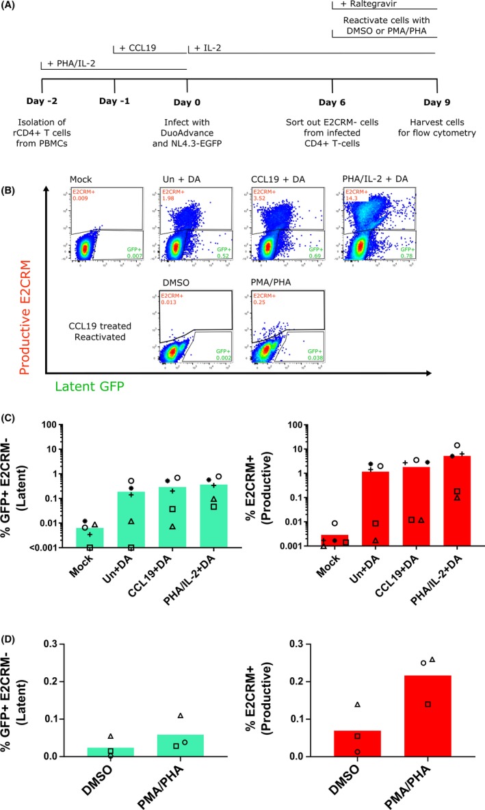
Figure 3.

E2 Crimson negative cells harbour inducible latent virus.
CD4+ T cells were isolated from PBMC and left resting (unstimulated, Un) or stimulated with either CCL19 or PHA/IL‐2 and then infected with either DuoAdvance (DA) virus or media only (mock) (A). After six days, expression of both fluorescent proteins was quantified by flow cytometry (B‐D). The E2 Crimson negative cell population (cells that were not present in the E2CRM+ gate in B top panel) was sorted from the CCL19‐treated samples six days after infection and then stimulated with either DMSO or PMA/PHA in the presence of 1 μM raltegravir and expression of both fluorescent proteins quantified by flow cytometry (D). Each symbol represents a different donor and the column represents the mean.
