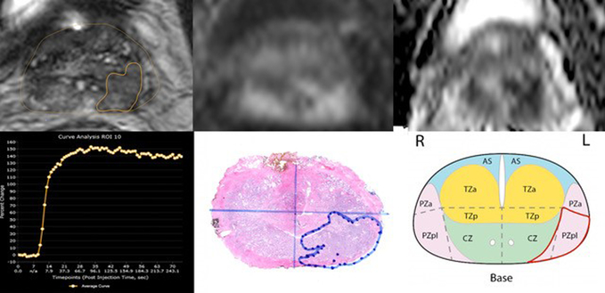
Fig. 2.
Multiparametric 3T MRI in a 66-year-old man with a history biopsy-proven PCa with PI-RADSv2 score of 4. (a) Axial T2-weighted MR image show peripheral zone lesion with 0.26 cm3 (b) DWI shows focal high intensity (c ) ADC map shows focal low intensity (d) DCE curve shows early and intense enhancement with immediate washout (e) Wholemount histopathology with maximum tumor diameter of 2 cm, GS 3+4 and tumor stage T2 (f) PI-RADSv2 sector map

