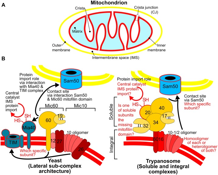
Fig 2. Conserved and diverged features of yeast and trypanosome MICOS.
(A) Basic mitochondrial architecture with relevant subcompartments indicated. (B) Comparison of MICOS complexes from yeast and trypanosomes. Color of outer and inner membrane phospholipid bilayers (double lines) as in (A). Subcomplex architecture indicated by brackets and color of component subunits. Only the numbers used in naming the MICOS subunits by the MicXX convention [22] are given. Conserved features of 2 complexes in black text with remaining uncertainties in red text. Extra-MICOS interactions indicated by back arrows pointing to interacting entity. Proteins involved in mitochondrial protein import in blue with red outline. CJ, crista junction; HS/SH, cysteine thiol side chain; IMS, intermembrane space; MICOS, mitochondrial contact site and cristae organization system; TIM, translocase of the inner membrane.

