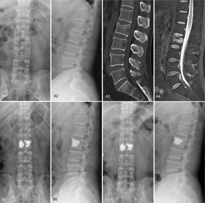
Figure 2.

A 57-year-old male diagnosed as L2 OVCF with “H” shaped cement filling pattern. Preoperative X-ray from coronal plane (a1) and sagittal plane (a2), CT image (a3), and MRI fat suppression image (a4). Postoperative X-ray from coronal plane (b1) and sagittal plane (b2) at 2-days follow-up. Postoperative X-ray from coronal plane (c1) and sagittal plane (c2) at 1-year follow-up.
