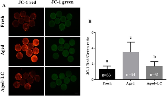
Fig. 3.

Effect of L-carnitine (LC) on the mitochondrial membrane potential (ΔΨm) of aged bovine oocytes in vitro. (A) Representative fluorescent images of JC-1-stained oocytes after in vitro aging. Scale bar: 200 µm, R = 3. (B) Quantification of JC-1 fluorescence intensity. Statistically significant differences are represented with different letters (P < 0.05).
