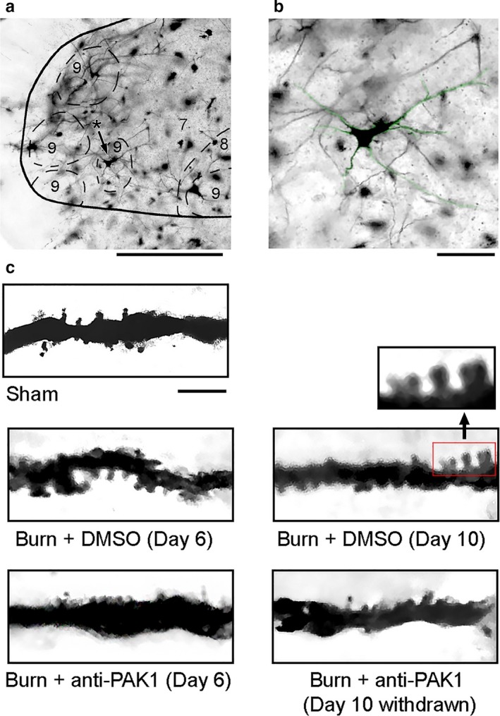
Figure 1.

Golgi‐stained tissue from the ventral spinal cord. (a) Coronal section of spinal cord showing a representative alpha‐motor neuron within lamina IX ventral horn (asterisk with black arrow). (b) Magnified motor neuron from panel a (green highlight). (c) Dendritic branch segments from alpha‐motor neurons sampled from each group: Sham, Burn + DMSO (Day 6), Burn + DMSO (Day 10), Burn + anti‐Pak1 (Day 6), and Burn + anti‐Pak1 (Day 10 withdrawal). Magnified view of multiple dendritic spines along a sampled dendritic branch is shown from the red box in panel c. Scale bars in A = 100 μm; B = 50 μm C = 10 μm
