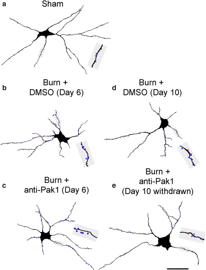
Figure 2.

Neurolucida digital reconstruction of alpha‐motor neurons. Digital rendering shows the morphology of a representative alpha‐motor neuron from (a) Sham, (b) Burn + DMSO (Day 6), (c) Burn + anti‐Pak1 (Day 6), (d) Burn + DMSO (Day 10), and (e) Burn + anti‐Pak1 (Day 10 withdrawal). Gray shaded boxes in each panel show a magnified view of a dendritic branch segment with thin (blue dots) and mushroom (red dots) dendritic spines. Scale bars a–c = 50 μm
