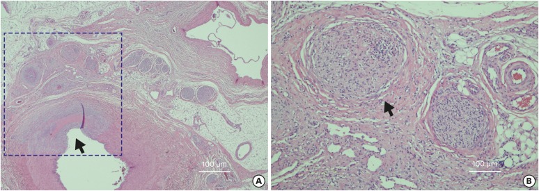
Figure 6. Histology of the renal artery. (A) H&E stain of the renal artery at low magnification shows degenerated renal artery and periarterial nerves. Renal artery shows loss of endothelium and medial thinning and fibrosis (black arrow). (B) High magnification view with H&E staining shows fibrosis of perineurium, marked pyknotic nuclei, eosinophilic change of nerve fibers, vacuolization, and inflammation (black arrow).
H&E = hematoxylin and eosin.

