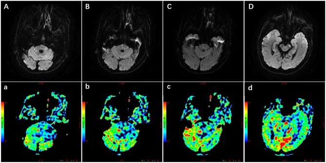
Figure 1.
Illustration of multiple infarctions and perfusion defects in the posterior circulation, which were detected by the diffusion-weighted imaging (DWI) (A–D) and perfusion weighted imaging (PWI) (a–d) respectively. The PWI images were the cerebral blood flow images corresponding to the DWI images. Scale was color coded (red, largest cerebral blood flow; blue, least cerebral blood flow). PWI images showed larger perfusion deficits including the brain stem, cerebellum, and occipital lobe.

