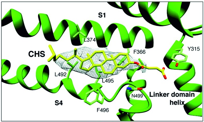
Figure 1.
A sterol binding pocket in the TRPC4DR structure. Electron cryo-microscopy structure of zebra fish TRPC4 (TRPC4DR) channel in its unliganded closed state, at an overall resolution of 3.6 Å. The transmembrane S1–S6 helices structure revealed that in the pre-S1 elbow domain inside the membrane, a cavity is formed with helices S1 and S4, in which a density corresponding to a sterol is formed. (Reproduced from Vinayagam et al. (2018) with permission from eLife.)

